
Table of Contents
Introduction
X-ray imaging is a technique for identifying and measuring photons or light particles with wavelengths in their part of the technique electromagnetic spectrum. X-rays are a type of radiation named electromagnetic waves. X-ray imaging generates pictures of the inside of your body. Scientists used it to help them understand the chemical properties and essential elements.
Many different x-ray imaging methods are used in many science and technology disciplines.
We can use these methods together or independently to create a more integrated image of degraded materials or objects.
Historically

Physicists and other scientists soon used his new technique. According to the SLAC National Accelerator Laboratory, between 1906 and 1908. British physicist Charles Barkla conducted research that concluded that X-rays could identify the diagnostic qualities of unique materials. And through his work, he received the Nobel Prize in Physics, but late in 1917
They used the x-ray spectrum to study the interaction of it’s radiation with atoms within the crystals. Their technique was called x-ray crystallography and became a standard on which scientists in subsequent years in the field of x-ray examination were based. They received the Nobel prize in physics in 1915.
How Is X-Ray Done?
When the atom is unstable or ejected by high-energy particles, electrons move from one energy level to another. When electrons converge, the element absorbs and releases high-energy x-ray photons in a way that characterizes the atoms that make up this particular chemical element.
Imaging measures these energy changes that allow scientists to identify elements and understand how atoms interact within different materials. There are two main methods of it’s imaging: wax and eds. Wax measures single-wavelength x-rays that will be reflecting through the crystal.
(Eds): measures the cytoid radiation emitted by stimulating electrons with a high-energy source of charged particles.
Multiple X-Ray Applications
We can use it’s imaging in many fields of science and technology, including archaeology, astronomy, engineering and health.
Anthropologists and archaeologists can discover hidden information about artefacts, and they still find them using X-ray imaging. For example, Lee Sharpe, assistant professor of chemistry at Grenell University in Iowa.
And his colleagues used a method called XRF to identify the origin of arrowheads made by prehistoric people in southwest North America, and published the results of its study in October 2018 in the Journal of Archaeology.
X-Ray Imaging Has Also Helped Astrophysicists
For example, researchers from Washington University in St. Louis plan to monitor X-rays from cosmic objects such as black holes to learn more about their properties. The team, led by Henric Krawczynskj, an experimental and theoretical astrophysicist, plans to launch its imaging called x-ray polarimeter.
Invisible spray antennas are only a few dozen thick nanometres. Still, they can transmit, and direct radio waves called xas imaging, helping us make sure thin materials are properly installing. And help us determine the conductivity of metals.
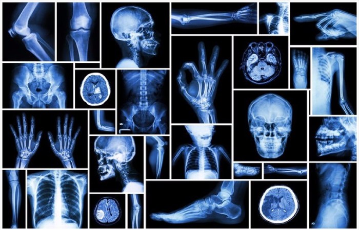
“High metal connectivity requires good antenna performance, so we have to keep a close eye on the material,” says gogotsi. We can also use imaging in many medical research and practice areas, such as modern ct Scan Machines.
Conclusion
Collecting X-ray absorption spectra during CT scans – by photonic counting or spectroscopy – can provide more detailed information about what happens inside the body, with radiation doses lower than X-rays and less or less needed to use heterogeneous substances (dyes). According to the Director of the Department of Radiology, Imaging and Science at Emory University in Georgia, Phuong-Anh T.Duong.